APEX — Arrhythmia Pattern Explainer

Clinical decision-support tool that reads 12-lead ECGs, detects abnormalities across 71 diagnostic categories, and generates grounded plain-English explanations with confidence-based review gating.

Source on GitHub Model card Python · PyTorch · Gradio
0.920macro-AUROC
71diagnostic categories
6 msinference latency
−97%calibration error

What the model produces

The figures and report cards below are real, unmodified output from the APEX pipeline — not mockups. This page is a static showcase; the interactive Gradio dashboard runs locally with make ui.
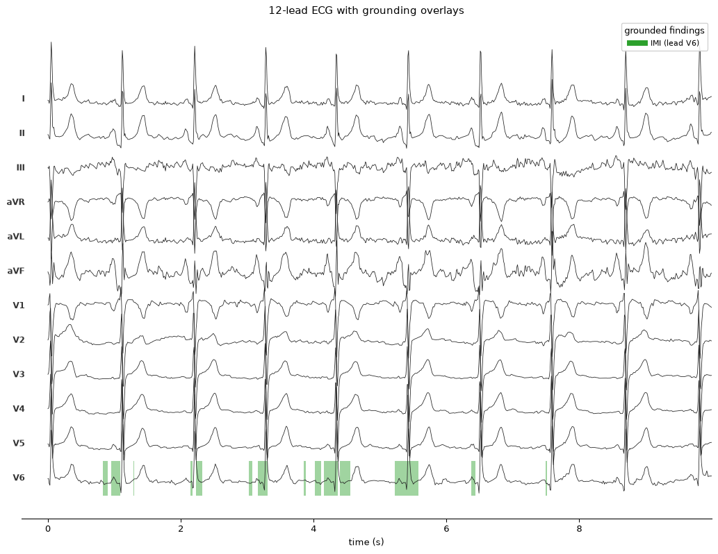
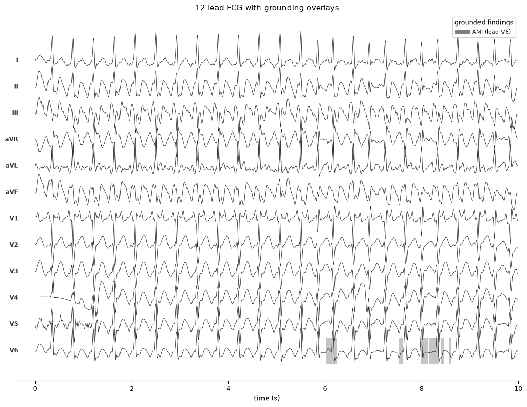
12-lead ECG with per-finding saliency overlays highlighting inferior leads II, III and aVF
Case 1 — inferior myocardial infarction. 12-lead waveform with per-finding grounding overlays; each colour maps to one detected finding.
ECG digitized from a photograph of a paper ECG, with saliency overlays
Case 2 — atrial fibrillation, digitized from a photograph of a paper ECG rather than a signal file.

Generated reports

Each report carries per-finding confidence, lead attribution, review flags, and a consistency check between the written explanation and the detector’s own saliency. Note the grounding conflict flag in the first card — the system reports when its explanation cites a lead that its saliency does not support.

🟡 Review recommended
One or more findings need clinician confirmation before acting.

Findings (4)

SR — sinus rhythm
78%  
ABQRS — abnormal QRS
93%  
IMI — inferior myocardial infarction● REVIEW
98%   leads II, III, aVF
grounding conflict
QWAVE — Q waves present● REVIEW
59%  
low confidence

Impression

Findings consistent with sinus rhythm. Inferior myocardial infarction.

Full report

Findings:
Regular rhythm with normal P waves preceding each QRS. Abnormal QRS morphology. Pathological Q waves in the inferior (II, III, and aVF) leads.

Impression:
Findings consistent with sinus rhythm. Inferior myocardial infarction.
✓ explanation consistent with detector
Decision support only — verify against the full clinical picture.
🟡 Review recommended
One or more findings need clinician confirmation before acting.

Findings (10)

AFIB — atrial fibrillation
94%  
AFLT — atrial flutter
100%  
PSVT — paroxysmal supraventricular tachycardia
86%  
SVARR — supraventricular arrhythmia
85%  
SVTAC — supraventricular tachycardia
98%  
2AVB — second degree AV block
86%  
LVOLT — low QRS voltages in the frontal and horizontal leads● REVIEW
63%  
low confidence
NST_ — non-specific ST changes
86%  
ABQRS — abnormal QRS● REVIEW
59%  
low confidence
AMI — anterior myocardial infarction● REVIEW
58%   leads V2, V3, V4
low confidencegrounding conflict

Impression

Findings consistent with atrial fibrillation. Atrial flutter. Paroxysmal supraventricular tachycardia. Supraventricular arrhythmia. Supraventricular tachycardia. Second-degree AV block. Non-specific ST changes. Anterior myocardial infarction.

Full report

Findings:
Irregularly irregular rhythm without discernible P waves. Intermittent failure of P-wave conduction to the ventricles. Low QRS voltage in the frontal and precordial leads. Non-specific ST-segment changes. Abnormal QRS morphology. Pathological Q waves in the anterior (V2, V3, and V4) leads.

Impression:
Findings consistent with atrial fibrillation. Atrial flutter. Paroxysmal supraventricular tachycardia. Supraventricular arrhythmia. Supraventricular tachycardia. Second-degree AV block. Non-specific ST changes. Anterior myocardial infarction.
✓ explanation consistent with detector
Decision support only — verify against the full clinical picture.